Patient Education

Tinnitus
Tinnitus or ringing in the ear occurs when sound is perceived but not coming from an external source. This happens due to damaged ear cells in the cochlea. Tinnitus can sound like a high pitched ringing, humming, or roaring, While there is no cure for tinnitus, there are methods to help make the tinnitus less noticeable. New technologies are being developed and tested to find a solution to tinnitus.
Watch VideoHelpful Links
A collection of helpful and informative links to more information about hearing aid services, speech therapy, general and pediatric ENT, cochlear implants, allergy information, asthma information and information related to head & neck cancers.
Read More
Eustachian Tube Dysfunction
Eustachian tube Dysfunction is a complication with the eustachian tubes. These tubes connect the inner ear to the back of the throat. Eustachian tubes function to equalize pressure behind the eardrum. They can become inflamed with ear infections which causes fluid to collect behind the eardrum. Symptoms: Popping or cracking sounds in the ear, Ear congestion / fullness, Ear pain.
Watch Video
Acoustic Neuroma
An acoustic neuroma (aka vestibular schwannoma) is a benign growth on the hearing nerve. It can cause a hearing loss affecting the ear that the tumor is growing. These are diagnosed with MRI imaging after seeing an asymmetrical hearing loss on an audiogram. The causes of these tumor growths is unknown at this time. Acoustic neuromas are rare and occur in about 10: 100,000 cases of asymmetric hearing loss. Treatment includes annual audiograms for observation, surgery or gamma knife radiation.
Watch Video
Benign Paroxysmal Positional Vertigo
The inner ear is the balance center of the body. Sometimes due to infection or injury, patients may experience a spinning sensation called vertigo. Vertigo triggered by moving the head in a certain directions. This is called Benign Paroxysmal Positional Vertigo (BPPV). There are crystals located in the inner ear that maintain balance but if they are knocked out of place, patients may experience BPPV.
Watch Video
Cerumen
Cerumen or earwax is naturally occurring and functions to protect the ear and keep the ear canal lubricated. It does not need removed unless it inhibits hearing or causes pain. ENTs have special tools to remove cerumen safely. Q-tips are not recommended as they can push earwax further into the canal as well as the potential to damage the eardrum
Watch Video
Cholesteatoma
Cholesteatoma occurs when there is an accumulation of dead skin cells found in the middle ear, inner ear, and or the mastoid bone. This can cause difficulty hearing and infection. Typically, this is found in patients who have had a history of recurrent ear infections. Symptoms: Decreased hearing, Ear pain, Ear fullness, Chronic ear infections, Problems with balance. Treatment: Medications (Antibiotics, Ear Drops), Surgical Management (Removal of Cholesteatoma).
Watch Video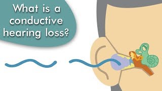
Conductive Hearing Loss
Conductive hearing loss occurs when there is an issue with sound waves reaching the middle and inner ear space. This can be due to mechanical obstruction like wax, foreign body, infection, or injury to the eardrum or hearing bones. These issues do not allow for sound waves to properly stimulate hair cells in the cochlea. Conductive hearing loss can be treated and the hearing loss is not always permanent. Treatment: Removal of obstruction. Surgery: (Tube surgery, cholesteatoma removal, prosthetic bone placement).
Watch Video
Meniere’s Disease
This disease is marked by dizziness, hearing loss, tinnitus, and ear congestion. These symptoms are not constant but occur during flare ups and may last up to 12 hours. Meniere’s Disease is caused by an abnormal amount of fluid in the semicircular canals. The cause is unknown. Symptoms: Dizziness, hearing loss, tinnitus, ear fullness. Treatments: Diet changes, medications, injections, surgery.
Watch Video
Myringotomies with Tubes
This procedure is typically performed on patients with recurrent ear infections. An incision is made in the tympanic membrane and the fluid is removed with a suction. Next, a small ventilation tube is placed in the eardrum. The tubes can last up to 18 months before they fall out. This is a very common treatment for babies and children with recurrent ear infections.
Watch Video
Otitis Media with Effusion
Otitis Media occurs when the middle ear space behind the tympanic membrane (eardrum) fills with fluid. The fluid can be clear or infected. Usually fluid behind the eardrum resolves by itself but if not, can be treated with antibiotics, steroids or by removing the fluid from the ear. If there is recurrence, your physician may advise tube placement for ventilation. This can be done in the office for adults or the operating room. Otitis media is the most common in the pediatric population due to immature eustachian tubes and being in daycare. Symptoms: Pain, Decreased hearing, Ears feeling clogged / congested. Treatment: Medications - antibiotics (sometimes steroids for adults). Surgery: Tubes placed in the ear canal.
Watch Video
Otosclerosis
Otosclerosis is a condition that affects the stapes bone in the inner ear. This bone is important in conducting sound. With otosclerosis, the bone overgrows, causing it to be less mobile. With less mobility, the cochlea is less responsive to stimuli, therefore decreasing hearing. Causes: Genetic. Treatments: Observation, Surgery.
Watch Video
Sensorineural Hearing Loss (SNHL)
Sensorineural hearing loss occurs when there is an issue with the cochlea hearing nerve. The cochlea contains small hair cells that receive and process sound waves. These signals are sent to the brain for interpretation. If the cochlea is damaged, these signals can’t be interpreted correctly by the brain. Some conditions that may cause SNHL include aging, prolonged exposure to loud sounds, congenital deformities, injury, or tumor. Treatment: Medication (Steroids if indicated). Surgery: Hearing Device Implants.
Watch Video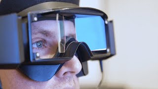
Videonystagamography (VNG)
This is a test that determines if the vertigo is caused by weakness in the right or left ear. This test places the patients in various positions and monitors eye movements with specialized goggles. Once the test is completed, an audiologist reads and interprets the report. Your physician will then go over the results with you and discuss treatment plans. The test typically lasts 1 hour long.
Watch Video
